UW Research Shows Camera in a Pill Can Offer Cheaper, Easier Window on Your Insides
Here are some highlights from an interesting research news as reported by the University of Washington (UW) Office of News and Information:
What if swallowing a pill with a camera could detect the earliest signs of cancer? The tiny camera is designed to take high-quality, color pictures in confined spaces. Such a device could find warning signs of esophageal cancer, the fastest growing cancer in the United States.
A fundamentally new design has created a smaller endoscope that is more comfortable for the patient and cheaper to use than current technology. Its first use on a human, scanning for early signs of esophageal cancer, will be reported in an upcoming issue of IEEE Transactions on Biomedical Engineering.
An endoscope is a flexible camera that travels into the body’s cavities to directly investigate the digestive tract, colon or throat. Most of today’s endoscopes capture the image using a traditional approach where each part of the camera captures a different section of the image. These tools are long, flexible cords about 9 mm wide, about the width of a human fingernail. Because the cord is so wide patients must be sedated during the scan.
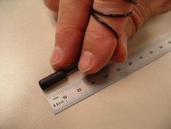
The scanning endoscope developed at the UW is fundamentally different. It consists of just a single optical fiber for illumination and six fibers for collecting light, all encased in a pill. Seibel acted as the human volunteer in the first test of the UW device. He reports that it felt like swallowing a regular pill, and the tether, which is 1.4 mm wide, did not bother him.
Once swallowed, an electric current flowing through the UW endoscope causes the fiber to bounce back and forth so that its lone electronic eye sees the whole scene, one pixel at a time. At the same time the fiber spins and its tip projects red, green and blue laser light. The image processing then combines all this information to create a two-dimensional color picture.
In the tested model the fiber swings 5,000 times per second, creating 15 color pictures per second. The resolution is better than 100 microns, or more than 500 lines per inch. Although conventional endoscopes produce images at higher resolution, the tethered-capsule endoscope is designed specifically for low-cost screening.
Using the scanning device is cheap because it’s so small it doesn’t require anesthesia and sedation, which increase the cost of the traditional procedure.
Click here for the complete article.